 After stroke, brain tumors are the most common cause of death from neurological diseases in the U.S., with 2% of all cancer deaths among adults attributable to primary malignant brain tumors. The incidence rate peaks in the seventh and eighth decades of adult life, and jumps by 500% in patients over age 85. Gliomas and meningiomas constitute the overwhelming majority of the pathological subtypes.
Clinical Presentation
To use a well-known real estate dictum, the clinical presentation of most primary brain tumors is dependent upon “location, location, location.” Although tumor-specific symptomatology can occur, most of the signs and symptoms of brain tumors are a function of the brain structures involved. A keen clinician will keep in mind the redundancy mechanisms built into the central nervous system (CNS) when examining a patient. Such redundancy can easily confound the clinical assessment, thus making imaging modalities, such as MR, powerful tools for identifying the exact location of a suspected lesion. At the same time, many brain tumors can present with completely nonspecific symptomatology, such as headaches. Headaches related to brain tumors may be related to hydrocephalus, hemorrhage, or direct efferent fiber irritation.
MRI Experience
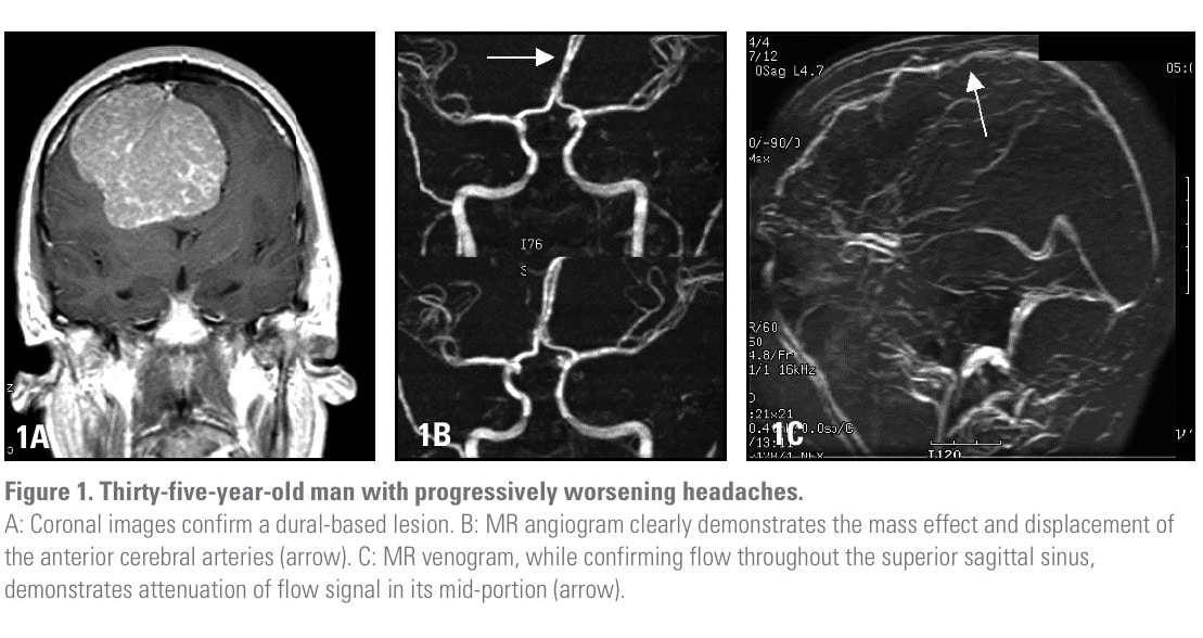
Contrast-enhanced MRI has revolutionized the imaging of intracranial neoplastic diseases. The exquisite soft-tissue detail combined with multiplanar imaging makes MR the imaging modality of choice in the assessment of brain tumors. The absence of ionizing radiation in MR eliminates concerns of radiation exposure in young patients and alleviates the anxiety related to repeated scans for close tumor follow-up. Even peritumoral arterial and venous structures can be assessed noninvasively with the addition of MR angiography and venography. (Figure 1)
A patient’s presenting symptoms combined with a clinician’s presumptive diagnosis helps tailor the MR examination. By adapting the imaging protocol to this pretest information, the specificity and sensitivity of MR is greatly enhanced. Some examples of such adaptations are reviewed below.
♦ Visual Disturbances
When symptomatology and physical examination direct concerns at the chiasmatic and prechiasmatic visual pathways, a directed orbital…
♦ Tinnitus and Hearing Loss
Objective tinnitus is a common symptom of vestibular schwannoma. When a clinician suspects the diagnosis of a vestibular schwannoma (acoustic neuroma)…
♦ Seizures
Given its innumerable etiologic factors, the initial imaging of patients with seizures needs to maintain a relatively…
♦ Headaches
When faced with nonspecific symptoms, MR examination of the brain can help exclude numerous etiologic factors…
Benign Tumors
• Meningiomas. Ninety percent of meningiomas are benign and most often found in middle-aged to elderly women. Although meningiomas can develop wherever leptomeningeal tissue is found, they have a proclivity for certain locations, such as the parasagittal dura, the convexities, and the sphenoid wings. Meningiomas are most commonly seen in the fifth and sixth decades, with a female-to-male ratio of 2:1. At all other ages, the ratio is closer to unity.

• Schwannomas are benign tumors derived from schwann cells, which provide myelin coverage to periphera…
• Trigeminal and facial schwannomas are a distant second and third in frequency. These lesions are sharply…
• Choroid plexus papillomas (CPP) are rare lesions with a propensity for the pediatric population…
• Arachnoid cysts, colloid cysts, and germ cell cysts represent the most common benign intracranial cystic…
• Epidermoid and dermoid cysts are both germ cell lesions arising from ectodermal elements displaced during…
Malignant Tumors
• Astrocytomas represent the most common form of malignant primary brain tumor. They share a common ancestral cellular origin with the oligodendroglia, the ependyma, and the choroid plexus…
• Oligodendrogliomas share a common cell lineage with astrocytomas. They occur most frequently in the fifth and sixth decades of life and are rare in…
• Ependymomas, in contradistinction to oligodendrogliomas, are primarily a tumor…
• Ganglion cell tumors are composed of mature neural elements. Admixture of neural and glial…
• Medulloblastomas. Over two-thirds of medulloblastomas occur in…
• Cerebral neuroblastomas tend to occur in the pediatric population…
• Primary brain lymphoma. Until recent years, primary brain lymphoma was a relatively rare neoplasm…
Advanced MRI Techniques
Recently, new MRI techniques have become available for the evaluation of parenchymal tumors…
- Diffusion tensor MR imaging with fiber tractography is a powerful method for in vivo…
- BOLD fMRI. Analysis of blood oxygen level-dependent functional MR imaging…
- Perfusion imaging has proved useful in the evaluation of primary tumors….
- MR spectroscopy has developed into a routine part of the MR armamentarium…
Unfortunately, the characteristic spectra of metastatic disease makes differentiation from primary tumors rather difficult…
Conclusion
Check out Spinal Cord Lesions by Gordon Sze at the Apple iBooks Store.
For more neurology-related articles click here.
