 The shoulder, or glenohumeral joint, is the most mobile but least stable joint in the human body. Disorders of the shoulder that result in chronic pain are common in both older and younger patients, particularly in those who participate in sports that involve frequent overhead motions, such as swimming, tennis, baseball, and gymnastics.
The shoulder can be affected by a variety of disorders, including arthritis, bursitis, acute trauma, abnormalities of the biceps tendon, neurologic disorders, frozen shoulder, tumor, and infection. Pathology involving the rotator cuff is one of the most common causes of shoulder problems.
Anatomy
Knowledge of the anatomy of the cuff is helpful for understanding the pathophysiology of rotator cuff disease. Composed of a group of muscles and tendons that surround the humeral head and glenohumeral joint, the cuff facilitates movement of the humeral head and stabilizes the head within the joint during motion.
Rotator Cuff Pathology
Pathologic disorders of the cuff include calcifying tendinopathy, tendinopathy with or without associated subacromial bursitis, and partial- or full-thickness tears. An uncommon but important complication of cuff disease..
Impingement
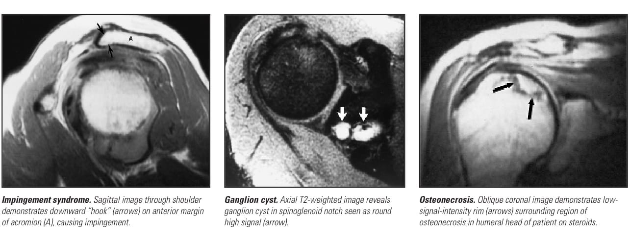
Impingement is generally seen in middle-aged and older individuals. It is often caused by spurs on the undersurface of the acromion or acromioclavicular joint. Other causes of impingement include…
Degeneration
Intrinsic degeneration of the rotator cuff can occur in the absence of impingement. In older patients, this is thought to result from vascular…
Tears
Cuff tendon tears tend to be chronic rather than acute. Patients usually do not recall an acute injury and may or may not have had a prolonged history…
Clinical Exam
Pain is the most common symptom associated with rotator cuff disease, although patients may also present with instability, stiffness, weakness, joint crepitus, deformity…
Treatment Options
Surgery is performed if conservative management is unsuccessful; it is also indicated in cases of acute cuff tears and when instability of the glenohumeral joint is associated with…
MRI–Clinical Experience
MRI is an accurate technique for detecting rotator cuff tears. Tears of the rotator cuff are more easily recognized…
• Partial-thickness tears can be seen on the articular or bursal cuff surface or may be completely enclosed…
Detection of the uncommon undersurface partial-thickness tears resulting from glenoid impingement can be improved by the use of MR arthrography. This is a procedure in which dilute MR contrast material is injected into the joint prior to the MR examination. It has been used primarily for the evaluation of labral abnormalities. • Impingement. MRI can depict many disorders that cause narrowing of the subacromial space, including spur formation beneath the acromion and acromioclavicular …
• Other disorders. MRI can also diagnose disorders other than rotator cuff disease that may be causing a patient’s shoulder pain…
Discussion
Rotator cuff disease is one of the most common causes of shoulder pain. While the clinical evaluation of shoulder pain often leads to an accurate diagnosis, the results can sometimes be misleading or insufficient to indicate appropriate…
- Plain films are very sensitive for detecting calcifications within the rotator cuff tendons….
- Arthrography is an accurate technique for detecting full-thickness cuff tears.4 It is relatively insensitive…
- Sonography of the cuff has been advocated by several investigators who have found it to be highly…
Conclusion
This article was edited from Impingment and Rotator Cuff Disease by the same author. The complete unabridged text, along with all related images and references, is available at the Apple iBooks Store.
Check out MR Arthrography of the Shoulder by Douglas P. Beall for an in-depth look at current concepts and clinical applications. For other riveting articles on the subject of musculoskeletal and joint disease imaging click here.
REFERENCES
- van de Sande MA, de Groot JH, Rozing PM. Clinical implications of rotator cuff degeneration in the rheumatic shoulder. Arthritis Rheum. Mar 15 2008;59(3):317-24.
- Corpus KT, Camp CL, Dines DM, et al. Evaluation and treatment of internal impingement of the shoulder in overhead athletes. World J Orthop. 2016 Dec 18;7(12):776-784.
- Lee YS, Jeong JY, Park CD, et al. Evaluation of the Risk Factors for a Rotator Cuff Retear After Repair Surgery. Am J Sports Med. 2017 Mar 1:363546517695234.
- Foti G, Avanzi P, Mantovani W, et al. MR arthrography of the shoulder: evaluation of isotropic 3D intermediate-weighted FSE and hybrid GRE T1-weighted sequences. Radiol Med. 2017 Feb 15.
