Lower back pain is a leading musculoskeletal cause of human impairment and disability. The costs – healthcare, physical therapy, lost work time/productivity – attributed to low back pain range from $16 to $90 billion per year. Common causes of low back and/or radicular pain are lumbar strain, herniated nucleus pulposus, lumbar spondylosis, tumor, and infection. Since MRI became available in the mid-1980s, evaluation of the lumbar spine has changed dramatically.
MRI – Clinical Experience
With its exceptional soft-tissue resolution, MR is the preferred imaging modality for intervertebral disc-related conditions. Disc desiccation at all stages is easily identified, and intervertebral disc protrusion is well visualized. Because of the high prevalence of spinal pathology in the asymptomatic population, the practitioner must be careful to correlate the results of imaging studies with the patient’s clinical examination.
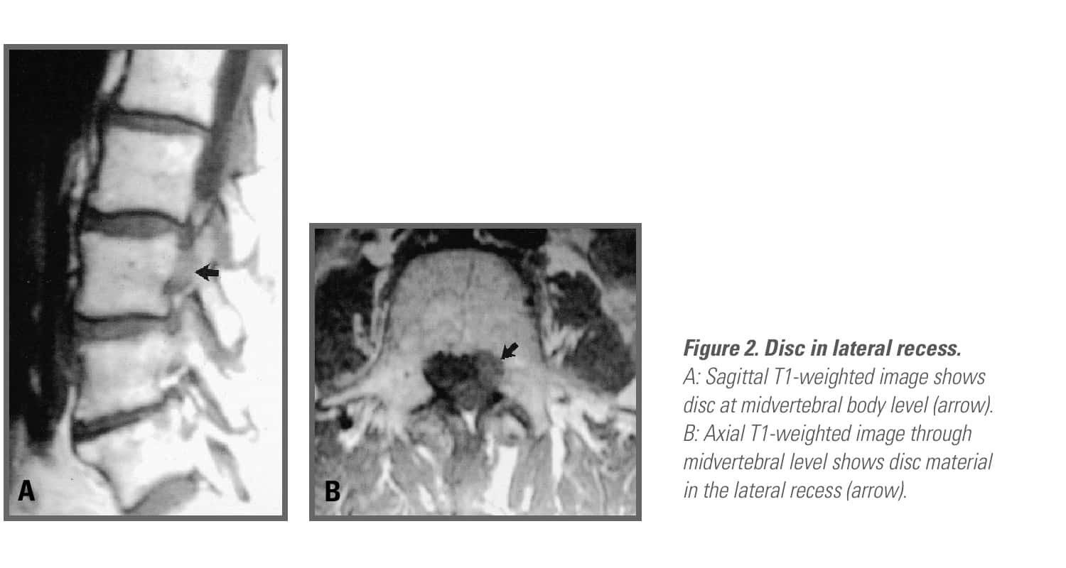
State-of-the-art imaging includes sagittal T1- and T2-weighted imaging. Contiguous axial imaging through the lumbar spine is necessary with T1 and T2 weighting. If evaluation in the axial plane is limited to the intervertebral disc space, then such entities as a free fragment of disc material that has migrated away from the disc space, pars interarticularis defect, and intraspinal synovial cysts can easily be overlooked…
Anatomical Types
Information concerning the location and type of pain, gathered from history and clinical examination, should guide the physician in planning the MRI evaluation.
• Lumbosacral Strain
Often the lower back is plagued by conditions such as strain of the lumbar soft tissues that result from…
• Herniated Nucleus Pulposus
Herniated nucleus pulposus (HNP) is one of the most common causes of sciatica, with a lifetime prevalence of approximately…
Guidelines for Using MRI in Lower Back Pain
MRI is an excellent modality for evaluation of radicular pain of lumbosacral origin. The ideal time to order further imaging is when the results will change treatment interventions. Following is a classification of disc ruptures:
- Bulge: circumferential symmetric extension beyond the interspace…
- Protrusion: focal or asymmetric extension of the disc beyond the interspace… and
- Extrusion: more extreme extension of the disc beyond the interspace…
MRI is not a screening test and should be used only to confirm the information obtained from a comprehensive history and physical examination. It should be reserved for certain clinical scenarios:
- When more invasive interventions such as spinal…
- When the etiology of the radicular pain remains…
- With cauda equina syndrome.
- In cases of suspicion for underlying…

• Lumbar Spondylosis
Lumbar spondylosis (arthritis) and spinal stenosis result from a progressive, noninflammatory arthritis of the lumbar facet joints…
Common objective findings in lumbar stenosis include radicular pain, dermatomal sensory loss, and…2
• Spinal Infections
Spinal infections are an uncommon but serious cause of low back pain. The vertebral body and disc can…
• Epidural Hematoma
In the cervical spine, an epidural hematoma requires emergent…
• Spinal Tumors
Tumors of the lumbar spine, either benign or malignant, are uncommon causes of backache, but these conditions…
• Fractures
Fractures, such as sacral insufficiency and sacral stress fractures, can also…
Discussion and Conclusion
This article was edited from Low Back Pain by the same author. The complete unabridged version, including all of its images and references, is available at the Apple iBooks Store. See other musculoskeletal/joint articles here.
REFERENCES
- Yong XZ, Sutherland T. Making sense of MRI of the lumbar spine. Aust Fam Physician. 2012 Nov; 41(11):887-90.
- Berg L, Hellum C, Gjertsen O, et al. Do more MRI findings imply worse disability or more intense low back pain? A cross-sectional study of candidates for lumbar disc prosthesis. Skeletal Radiol. 2013 Nov;42(11):1593-602.
- Suthar P, Patel R, Mehta C, Patel N. MRI evaluation of lumbar disc degenerative disease. J Clin Diagn Res. 2015 Apr;9(4):TC04-9.
- Talbott JF, Narvid J, Chazen JL, et al. An Imaging-Based Approach to Spinal Cord Infection. Semin Ultrasound CT MR. 2016 Oct;37(5):411-30.

